This content is for informational and educational purposes only. Always consult a qualified healthcare provider.
Last Updated on July 1, 2026 by Grace Oluchi
Neuroimaging is a branch of science that uses lots of technologies to produce images of the brain or other parts of the nervous system in a noninvasive way. Neuroimaging can help us study the structure and function of the brain, as well as the changes that occur in the brain due to diseases, injuries, or other factors. Neuroimaging can also help to diagnose and treat many neurological and psychiatric disorders, as well as monitor the effects of interventions such as drugs, surgery, or stimulation.
Neuroimaging can be divided into two broad categories:
- Structural.
- Functional.
- Structural neuroimaging is used to visualize and measure the anatomy and morphology of the brain, such as the size, shape, and location of different brain regions, tissues, or cells.
- Functional neuroimaging is used to assess the activity and metabolism of the brain, such as the blood flow, oxygen consumption, glucose uptake, or electrical signals of different brain regions, cells, or networks.
📋 Table of Contents
What Are the Main Types of Neuroimaging Techniques?
- Computed tomography (CT) scan: This technique uses X-rays to create cross-sectional images of the brain. It can show the bones, blood vessels, and soft tissues of the brain, as well as detect abnormalities such as tumors, bleeding, or fractures. The but is that, it also exposes the patient to radiation, which can have harmful effects in the long term. CT scan is also not very sensitive to small changes in the brain structure or function.
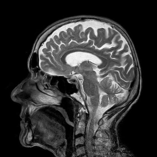
- Magnetic resonance imaging (MRI) scan: This technique uses a strong magnetic field and radio waves to create detailed images of the brain. It can show the anatomy and morphology of the brain, as well as the <a href="https://medspurs.com/category/food-and-nutrition/” class=”il-link”>water content, blood flow, and chemical composition of different brain tissues. It can also detect abnormalities such as inflammation, infection, or damage. MRI scan does not use radiation, but it can be noisy, expensive, and time-consuming. MRI scan is also not suitable for patients with metal implants or devices, such as pacemakers or cochlear implants.
- Positron emission tomography (PET) scan: This technique uses a radioactive tracer that is injected into the bloodstream and taken up by the brain cells. The tracer emits positrons, which are detected by a special camera that creates images of the brain. PET scan can show the metabolism and function of the brain, such as the glucose uptake, oxygen consumption, or neurotransmitter activity of different brain regions or cells. It can also detect abnormalities such as tumors, infections, or degeneration. PET scan uses radiation, which can have harmful effects in the long term. PET scan is also expensive, invasive, and requires a cyclotron to produce the tracer.
- Functional magnetic resonance imaging (fMRI) scan: This technique uses the same principle as MRI scan, but it focuses on the changes in the blood oxygen level dependent (BOLD) signal that reflect the brain activity. fMRI scan can show the activation and connectivity of different brain regions or networks during various tasks or stimuli. It can also measure the resting state activity of the brain, which reflects the intrinsic function and organization of the brain. fMRI scan does not use radiation, but it can also be noisy, expensive, and time-consuming. fMRI scan is also affected by motion, breathing, or heart rate, which can introduce noise or artifacts in the images.
- Electroencephalography (EEG) scan: This technique uses electrodes attached to the scalp to measure the electrical activity of the brain. EEG scan can show the frequency, amplitude, and phase of the brain waves, which reflect the state and function of the brain. EEG scan can also detect abnormalities such as seizures, epilepsy, or brain death. EEG scan does not use radiation, but it can be affected by external noise, muscle activity, or eye movements, which can introduce noise or artifacts in the signals. EEG scan is also not very accurate in locating the source of the brain activity, as it suffers from the inverse problem.
- Magnetoencephalography (MEG) scan: This technique uses sensors placed around the head to measure the magnetic fields generated by the electrical activity of the brain. MEG scan can show the frequency, amplitude, and phase of the brain waves, which reflect the state and function of the brain. MEG scan can also detect abnormalities such as seizures, epilepsy, or brain tumors. MEG scan does not use radiation, but it requires a shielded room to block the external magnetic fields, which are expensive and scarce. MEG scan is also more accurate than EEG scan in locating the source of the brain activity, as it suffers less from the inverse problem.
- Near-infrared spectroscopy (NIRS) scan: This technique uses light sources and detectors placed on the scalp to measure the changes in the blood oxygenation and hemoglobin concentration in the brain. NIRS scan can show the activation and connectivity of different brain regions or networks during various tasks or stimuli. NIRS scan does not use radiation, but it can be affected by scalp blood flow, hair, or skin pigmentation, which can introduce noise or artifacts in the signals. NIRS scan is also not very deep in penetrating the brain tissue, as it can only reach the outer layer of the cortex.
What are Some of the Applications and Challenges of Neuroimaging?
- Neuroscience: Neuroimaging can help us understand how the brain works, how it develops, how it ages, and how it is affected by different factors, such as genes, environment, drugs, or diseases. Neuroimaging can also help us find out the neural basis of cognition, emotion, behavior, and consciousness.
- Neurology: Neuroimaging can help us diagnose and treat various neurological disorders, such as stroke, Alzheimer’s disease, Parkinson’s disease, multiple sclerosis, epilepsy, or brain tumors. Neuroimaging can also help us monitor the progression and outcome of these disorders, as well as the effects of interventions, such as drugs, surgery, or stimulation.
- Psychiatry: Neuroimaging can help us diagnose and treat various psychiatric disorders, such as <a href="https://medspurs.com/category/mental-health/” class=”il-link”><a href="https://medspurs.com/category/mental-<a href="https://medspurs.com/category/health/” class=”il-link”>health/” class=”il-link”>depression, schizophrenia, bipolar disorder, <a href="https://medspurs.com/category/mental-<a href="https://medspurs.com/category/health/” class=”il-link”>health/” class=”il-link”>anxiety, or autism. Neuroimaging can also help us understand the causes and mechanisms of these disorders, as well as the effects of interventions, such as drugs, psychotherapy, or stimulation.
- Education: Neuroimaging can help us study how the brain learns, remembers, and processes information. Neuroimaging can also help us design and test effective teaching and learning methods, as well as identify and support students with learning difficulties or disabilities.
- Law: Neuroimaging can help us assess the mental state, capacity, and responsibility of individuals involved in legal cases, such as criminals, witnesses, or victims. Neuroimaging can also help us detect deception, memory, or emotion in legal contexts, such as interrogations, trials, or negotiations.
- Ethics: Neuroimaging can help us find out the moral and social implications of using neuroimaging for various purposes, such as research, diagnosis, treatment, enhancement, or manipulation. Neuroimaging can also help us address the ethical issues and challenges that come from neuroimaging, such as privacy, consent, validity, reliability, or interpretation.
Limitations That Should Be Addressed.
- Technical: Neuroimaging techniques are usually difficult, expensive, and time-consuming. They also need specialized equipment, expertise, and infrastructure. They also have different trade-offs, such as spatial resolution, temporal resolution, signal-to-noise ratio, or invasiveness. They also have lots of sources of error, noise, or artifact, such as motion, physiology, or environment.
- Methodological: Neuroimaging data are often large, multidimensional, and heterogeneous. They also require sophisticated processing, analysis, and interpretation. They also have various assumptions, models, and parameters that need to be validated and optimized. They also have various biases, confounds, or pitfalls that need to be controlled and corrected.
- Theoretical: Neuroimaging findings are often correlational, not causal. They also require integration and synthesis with other sources of information, such as behavior, genetics, or pharmacology. They also require conceptual and theoretical frameworks and hypotheses that guide and explain the neuroimaging results. They also require generalization and replication across different populations, settings, and conditions.
- Practical: Neuroimaging applications are often limited by the availability, accessibility, and affordability of neuroimaging resources and services. They also require collaboration and communication among various stakeholders, such as researchers, clinicians, educators, lawyers, or policymakers. They also require regulation and governance that ensure the quality, safety, and ethics of neuroimaging practices and products.
FAQs on Neuroimaging.
What is the difference between MRI and fMRI?
MRI stands for magnetic resonance imaging, which is a technique that uses a strong magnetic field and radio waves to create detailed images of the brain structure. fMRI stands for functional magnetic resonance imaging, which is a technique that uses the same principle as MRI, but focuses on the changes in the blood oxygen level dependent (BOLD) signal that reflect the brain activity.
What is the inverse problem in neuroimaging?
The inverse problem in neuroimaging is the challenge of inferring the source of the brain activity from the measured signals, such as EEG or MEG. The inverse problem is difficult because there are many possible sources that can produce the same signals, and there is no unique solution to the problem. So, the inverse problem requires making assumptions, models, and constraints to reduce the uncertainty.
What is the default mode network (DMN) in neuroimaging?
The default mode network (DMN) is a network of brain regions that are more active when the person is at rest, not engaged in any specific task, or thinking about themselves or others. The DMN includes the medial prefrontal cortex, the posterior cingulate cortex, the precuneus, the inferior parietal lobule, and the hippocampus. The DMN is thought to be involved in self-referential, introspective, and social processes, such as memory, imagination, empathy, and moral reasoning.
What is the blood-brain barrier (BBB) and how does it affect neuroimaging?
The blood-brain barrier (BBB) is a protective layer of cells that separates the blood vessels from the brain tissue. The BBB regulates the exchange of substances between the blood and the brain, and prevents the entry of harmful agents, such as toxins, pathogens, or drugs. The BBB affects neuroimaging because it limits the availability and delivery of contrast agents or tracers that are used to enhance the visibility or specificity of the brain images, such as in CT, PET, or MRI.
What is neurofeedback and how does it use neuroimaging?
Neurofeedback is a type of biofeedback that uses neuroimaging to monitor and modulate the brain activity of the person. Neurofeedback involves presenting the person with a real-time feedback of their brain activity, such as a visual, auditory, or tactile stimulus, and instructing them to change or control their brain activity in a desired way, such as increasing or decreasing the frequency of certain brain waves. Neurofeedback can use neuroimaging techniques such as EEG, fMRI, or NIRS to measure and display the brain activity.
What is multimodal neuroimaging and what are its advantages?
Multimodal neuroimaging is the combination of two or more neuroimaging techniques to study the brain structure and function in a complementary and synergistic way. Multimodal neuroimaging can overcome the limitations of each individual technique and provide more detailed and accurate information about the brain. For example, combining fMRI and EEG can provide both high spatial and temporal resolution of the brain activity, while combining PET and MRI can provide both metabolic and anatomical information of the brain.